How Dentists Diagnose and Treat Cavities?

Understanding Dental Cavities

Definition and Causes
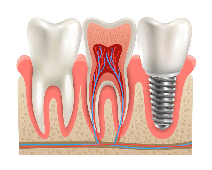
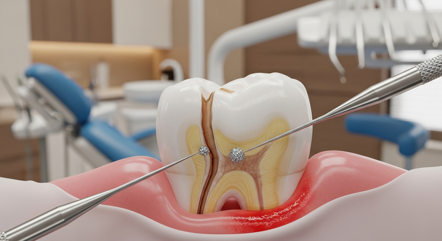
Cavities are decayed areas of teeth that develop into tiny openings or holes. The primary causes of cavities include bacteria in the mouth, frequent snacking, sipping sugary beverages, and inadequate oral hygiene. The three common types of cavities are:
Maintaining proper oral hygiene is essential for prevention. Regular brushing, flossing, and visiting the dentist can significantly reduce the risk of developing cavities. If left untreated, cavities can grow, affecting deeper layers of teeth and potentially leading to toothaches or infections (Mayo Clinic).
Risk Factors and Prevalence
Cavities can affect anyone with teeth, including children, teenagers, and older adults. Certain factors increase the risk of cavity formation, such as:
It is critical to understand these risk factors to implement effective preventive measures. The following table summarizes the prevalence of cavities in different age groups:
Age GroupPrevalence of CavitiesChildren (ages 2-11)42%Teens (ages 12-19)59%Adults (ages 20-64)91%Older Adults (65+)96%
Knowing how to prevent cavities is vital, as untreated cavities can lead to severe dental problems, including tooth loss. Regular dental check-ups and practicing good oral hygiene are crucial for ensuring dental health. For more information on the importance of regular visits to the dentist, check out our article on why you should see a dentist regularly?.
Diagnosing Cavities
Dentists employ various methods to diagnose cavities, ensuring that they can effectively identify and treat tooth decay at early stages. This section covers the primary techniques used in cavity detection: visual examination, radiographic techniques, and advanced diagnostic tools.
Visual Examination
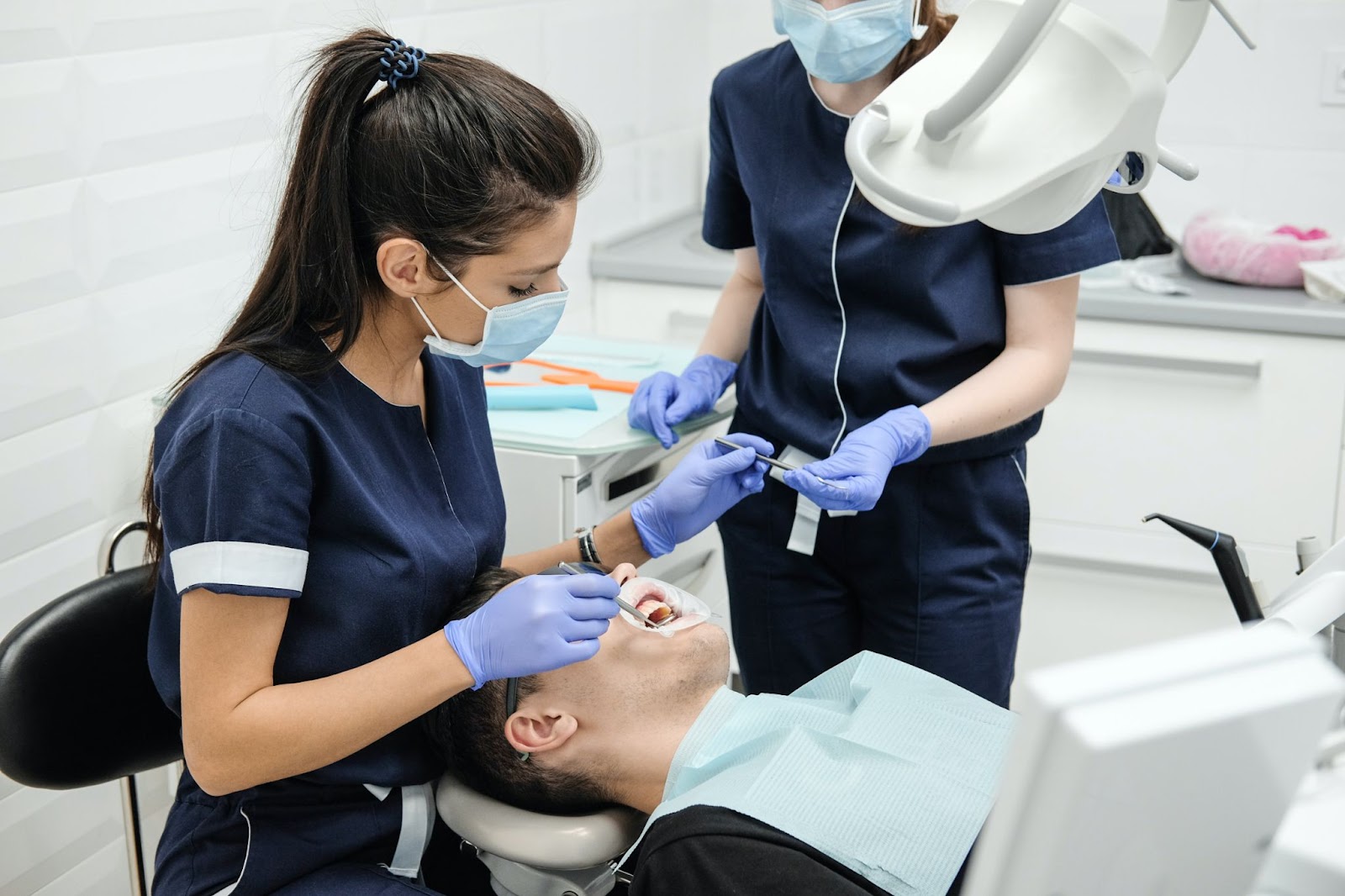
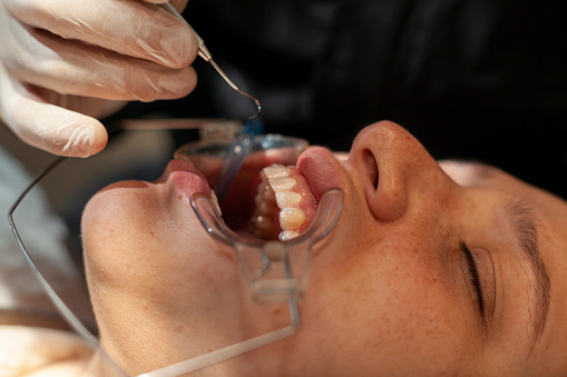
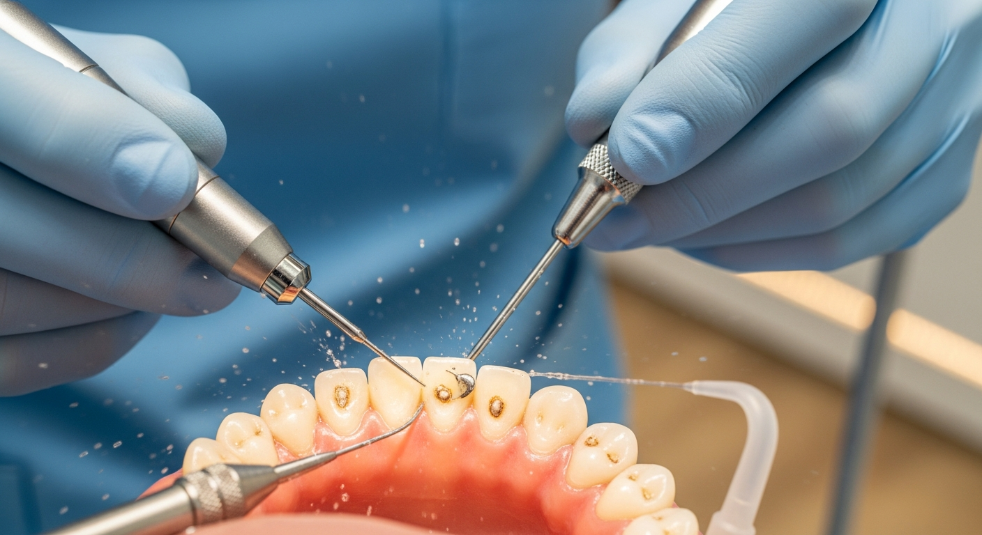
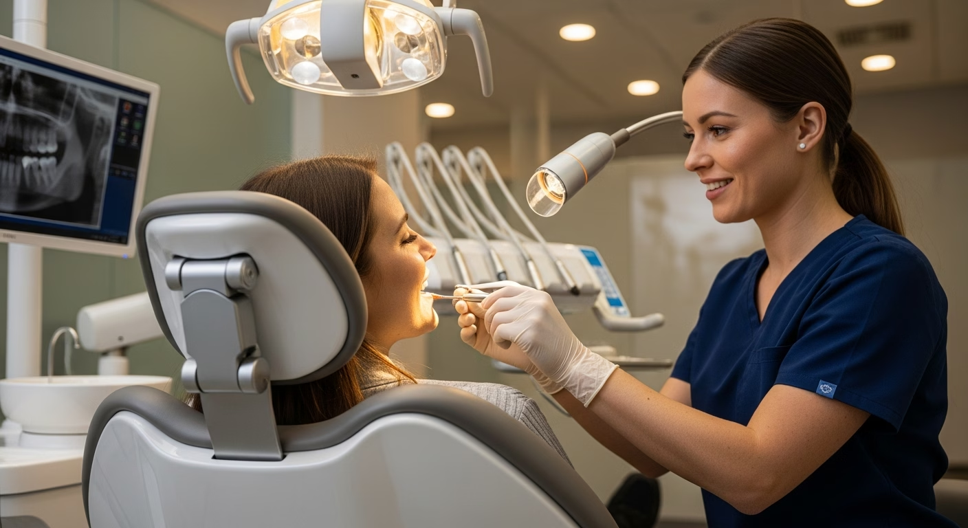
The first step in diagnosing dental cavities typically involves a visual examination. During this process, the dentist visually inspects the teeth for any visible signs of decay, discoloration, or damage. They will also check the gums and surrounding tissues for any abnormalities. According to the NCBI Bookshelf, this examination may include using a dental explorer to probe any suspicious areas of teeth.
AspectDescriptionVisibilitySigns of decay, cavities, or discolorationTechniquesUse of a dental explorer and mirror
Radiographic Techniques
If the visual examination suggests the presence of cavities, dentists often rely on radiographic techniques to obtain a more detailed view of the teeth. X-rays can help detect decay that is not visible to the naked eye, especially in areas between teeth. The NCBI Bookshelf emphasizes the importance of X-rays in identifying cavities below the surface of the enamel.
Type of X-rayPurposeBitewing X-raysDetect cavities between teethPeriapical X-raysExamine the entire tooth structure including the roots
Advanced Diagnostic Tools
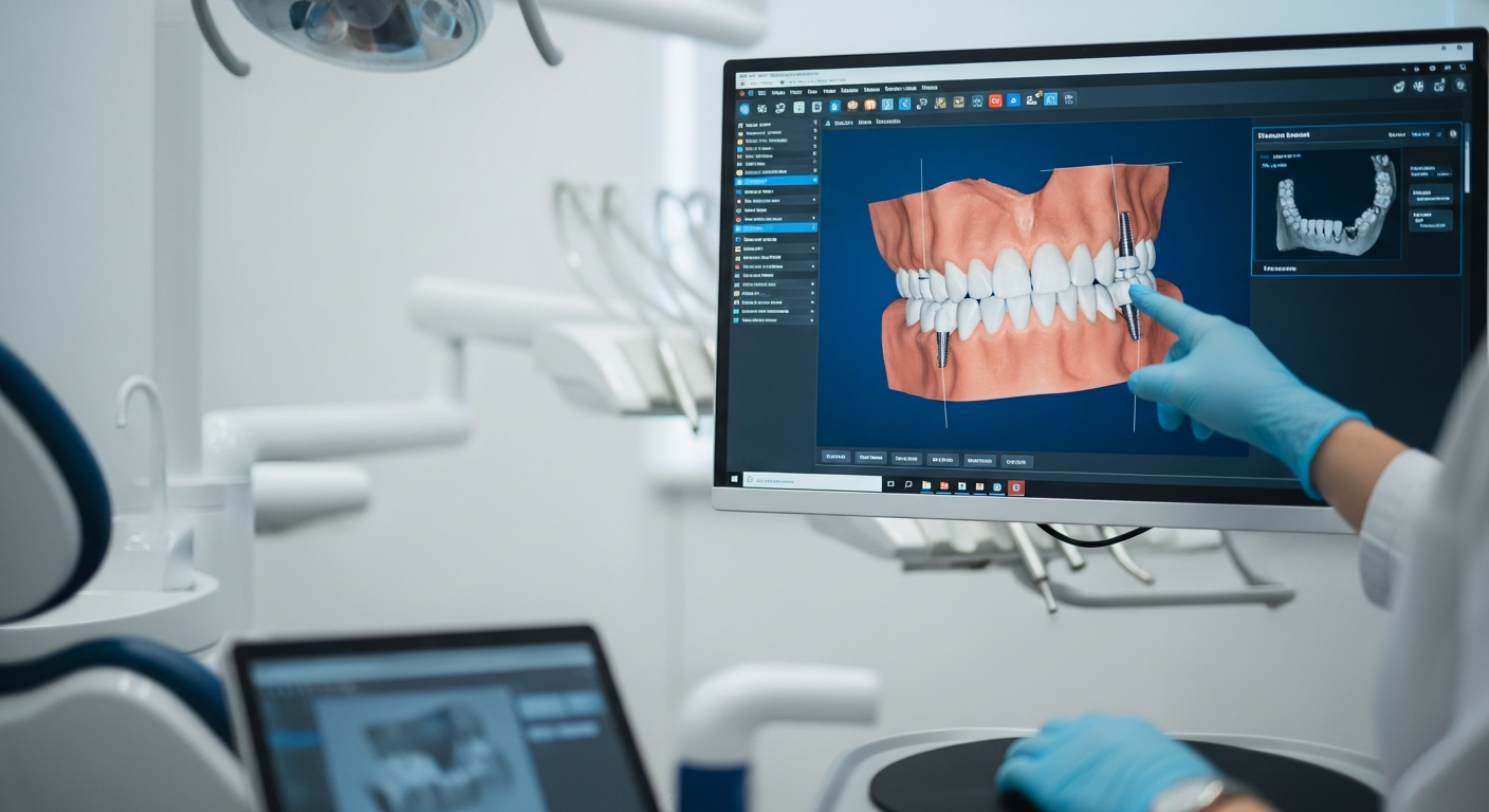
In addition to the traditional methods, dentists may use advanced diagnostic tools to enhance cavity detection capabilities. These tools may include laser technology and digital imaging, which can provide more precise information about tooth health. The use of diode lasers has been noted for its effectiveness in early cavity detection, as highlighted by both the NCBI Bookshelf and the Dental Health Society.
ToolDescriptionDiode lasersHelp in detecting cavities and assessing tooth healthDigital imagingProvides high-resolution images for better analysis
Each of these methods contributes to a comprehensive understanding of a patient’s dental health. By combining visual examinations, radiographic techniques, and advanced diagnostic tools, dentists can effectively diagnose cavities and determine the most appropriate treatment options. For more information on how dentists treat cavities, refer to our section on treatment options.
Treatment Options for Cavities
Once a dentist diagnoses a cavity, several treatment options are available depending on the severity of the decay. The most common treatments include dental fillings, crowns, root canal therapy, and tooth extraction as a last resort.
Dental Fillings
Dental fillings are the most prevalent treatment for moderate to severe cavities. During the procedure, the dentist removes decayed tissue from the tooth, creating a clean cavity. Then, they fill the prepared space with appropriate materials, which may include composite resin, gold, or silver, depending on the cavity's severity and location.
Filling MaterialDescriptionEstimated LongevityComposite ResinTooth-colored filling that blends with the tooth5-10 yearsGoldDurable and long-lasting, less aesthetic10-15 yearsSilver (Amalgam)Made from a mixture of metals, durable10-15 years
Crowns and Root Canal Therapy
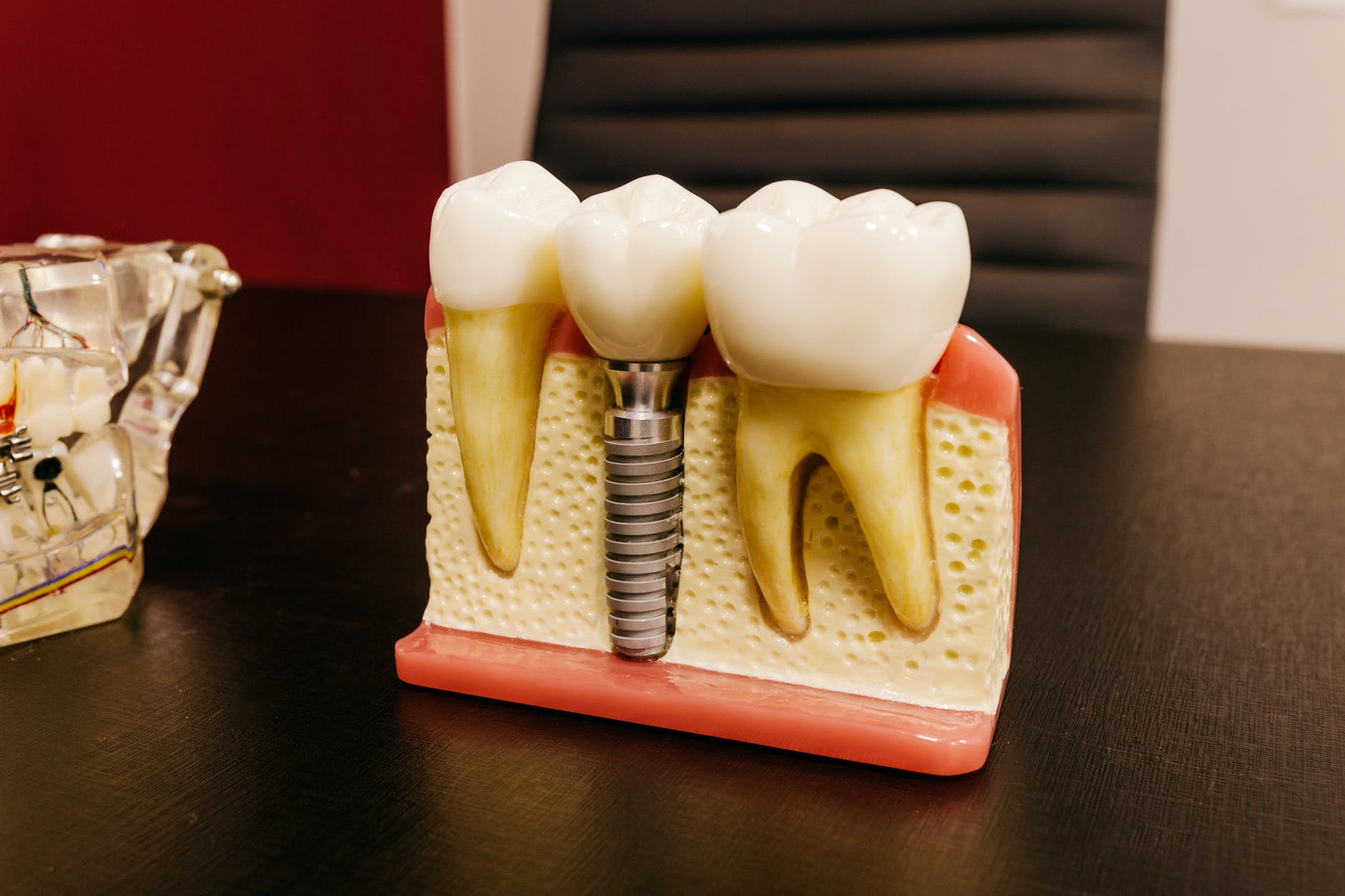
In cases where a filling alone cannot provide sufficient protection, especially if a large filling leaves the tooth vulnerable to cracking, crowns may be utilized. The dentist covers the repaired tooth with an alloy or porcelain crown to ensure protection.
Root canal therapy is another option for cavities that have significantly damaged the tooth nerves. When decay penetrates deep into the tooth, affecting the nerves, the dentist removes the damaged nerve and fills the area with an endodontic sealant. A crown is often placed over the affected tooth afterward to restore its strength and functionality (Oral-B).
Tooth Extraction as a Last Resort
Tooth extraction is considered a last resort in cavity treatments. It is explored when other treatment options are insufficient or when decay increases the risk of infection spreading to the jawbone. Simple extractions usually do not require incisions or general anesthesia, making them more straightforward procedures.
Understanding these treatment options helps individuals be better informed about how dentists diagnose and treat cavities. Regular dental check-ups are essential for early detection and intervention, significantly reducing the need for more invasive treatments.
Dental Filling Procedure
The dental filling procedure involves several critical steps to restore a tooth affected by decay. Here, the focus will be on assessment and material selection, anesthesia and cavity preparation, and filling placement and post-filling care.
Assessment and Material Selection
Before the filling procedure, the dentist performs a thorough assessment of the tooth and the cavity. The type of cavity can differ; for instance, occlusal cavities occur on chewing surfaces, while interproximal cavities are located between teeth (Oral-B).
The dentist selects the appropriate filling material, which can include:
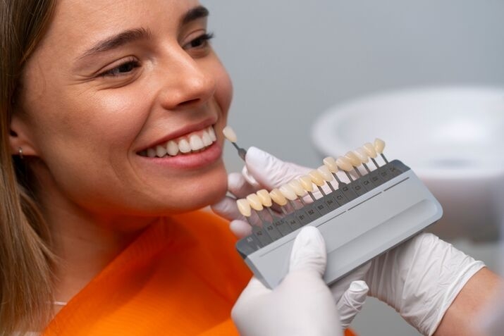
Filling MaterialDescriptionDirect Composite BondingTooth-colored material for aesthetic needsPorcelainDurable and excellent for cosmetic fillingsGlass IonomerReleases fluoride, suitable for childrenSilver AmalgamKnown for strength, often used in back teethGold InlaysHighly durable, utilized for restoration cases
The choice of material is influenced by various factors such as the cavity's location, the patient's medical history, aesthetic preferences, and financial considerations (News-Medical).
Anesthesia and Cavity Preparation
To ensure patient comfort, a local anesthetic is administered before beginning the filling process. This numbs the surrounding area of the affected tooth, making the procedure more comfortable (News-Medical).
After numbing, the dentist uses a dental drill to remove the decayed portion of the tooth. Following decay removal, the cavity is thoroughly cleaned to prevent infection and ensure proper bonding of the filling material.
Filling Placement and Post-Filling Care
Once the cavity is prepared, the selected filling material is applied. The dentist shapes it to match the contour of the tooth and ensures that it does not interfere with the patient's bite. Additional polishing is performed to enhance the smoothness of the filling (Dentistry of West Bend).
Post-filling care is essential to ensure proper healing and longevity of the filling. Patients are typically advised to avoid extremely hot or cold foods for a few days. They may also receive instruction on maintaining oral hygiene and monitoring any unusual sensations, which could indicate the need for a follow-up visit.
Understanding these steps in the dental filling process highlights how dentists effectively diagnose and treat cavities. For more information on cavity management, visit our section on how dentists diagnose and treat cavities?.
Importance of Regular Dental Check-ups
Regular dental visits are vital for maintaining oral health and preventing more complex issues in the future. By understanding the significance of preventive measures and the benefits of early detection and intervention, individuals can take proactive steps toward optimal dental care.
Preventive Measures
Preventive care involves actions taken to prevent dental issues before they occur. Regular check-ups enable dentists to perform cleanings, remove plaque, and conduct thorough assessments of dental health, which are essential for maintaining a healthy smile. Routine visits allow for the following preventive measures:
These measures are shown to significantly reduce the likelihood of cavities and gum disease, contributing to a healthier mouth.
Early Detection and Intervention
One of the critical advantages of regular dental check-ups is the opportunity for early diagnosis of problems. Detecting issues such as cavities in their initial stages can prevent the need for more extensive treatments in the future (Mayo Clinic). Early intervention can include:
Visiting the dentist regularly is essential for maintaining good oral health, as it allows for timely intervention when problems arise (Dentistry of West Bend). Regular check-ups can significantly affect long-term dental health and enhance overall well-being.
For those interested in enhancing their dental care experience, learning more about how to choose a cosmetic dentist near me? can provide valuable insights.
Innovative Technologies in Cavity Detection
With advancements in dental technologies, diagnosing cavities has become more precise and efficient. One of the innovative tools gaining prominence is the diode laser.
Diode Lasers in Dentistry
Diode lasers are increasingly utilized by dental professionals for cavity detection, as well as for treating various oral health issues such as gingivitis, laser gum reshaping, and teeth whitening (Dental Health Society). These lasers work by measuring the density of teeth through a technique known as transillumination. In this process, light is shined on the tooth, and the way this light reflects back can help distinguish healthy enamel from areas affected by decay.
Role in Early Detection
One of the significant advantages of diode lasers is their capability to identify initial signs of decay earlier than traditional x-rays. The reflection of light from decayed areas is registered as a numerical reading, with higher numbers indicating more severe decay (Dental Health Society). This early detection may allow dentists to intervene before dramatic procedures like drilling or anesthetic are needed, potentially preserving more of the natural tooth structure.
Detection MethodPotential UseSensitivityDiode LasersEarly cavity detectionSensitive but potentially leads to unnecessary fillingsTraditional X-raysDetect decay, especially between teethEffective for deep decay detection
Benefits and Limitations
While diode lasers offer numerous benefits, there are also limitations to their use. They are adept at identifying cavities on the chewing surfaces of teeth but may be less effective at detecting decay located between teeth or deeper within a tooth. For comprehensive examinations, most dentists opt to use a combination of tools, including both diode lasers and traditional x-rays, to ensure thorough cavity detection (Dental Health Society).
Additionally, diode lasers can occasionally be overly sensitive, which might lead to unnecessary treatments for small spots detected. To assess whether treatment is necessary, dentists may monitor these areas over time.
By integrating innovative technologies like diode lasers into their practice, dentists are better equipped to provide more accurate diagnoses and improved treatment options. For further information on how dentists diagnose cavities, visit our section on how dentists diagnose and treat cavities?.




















.avif)







.jpg)


.avif)


.jpg)


